
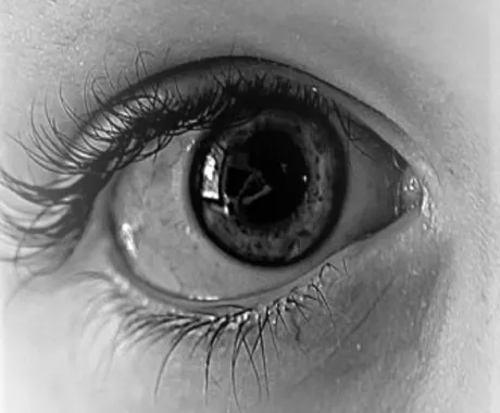
A dilated eye exam is required to gain a good view of the inside of your eyes so that an assessment can be made of your overall eye health.
This involves instilling diagnostic medication in the form of eye drops so that your pupils will get bigger, allowing a better view of your lenses, retina, macula, optic nerve, and blood vessels.
The drops take about 15–30 minutes to kick in before your pupils open up. You will be left with blurry vision and some glare for 2–3 hours afterwards, so it is best to bring sunglasses and arrange a ride or use public transport.
This is usually conducted at a follow-up examination as it requires more time. If you’d like it during your initial examination, please let me know ahead of time so I can ensure you have an extended appointment.
Regular eye exams prevent blindness
Many serious eye conditions progress silently, with no pain or obvious symptoms until significant damage has occurred.
- 50% of Australians with glaucoma are undiagnosed due to a lack of regular eye exams. Blindness from glaucoma is preventable if detected early.
- The myopia epidemic causes irreversible vision loss. Myopic maculopathy is the leading cause of blindness in the Asian population, and it cannot be corrected with glasses.
- Macular degeneration (AMD) is responsible for 50% of blindness in Australia. Symptoms are rare in the early stages and often missed because they tend to start in one eye only.
- All diabetics need regular dilated eye examinations at least once every 2 years. Diabetic eye disease is the number one cause of preventable blindness in the working-age population in Australia.
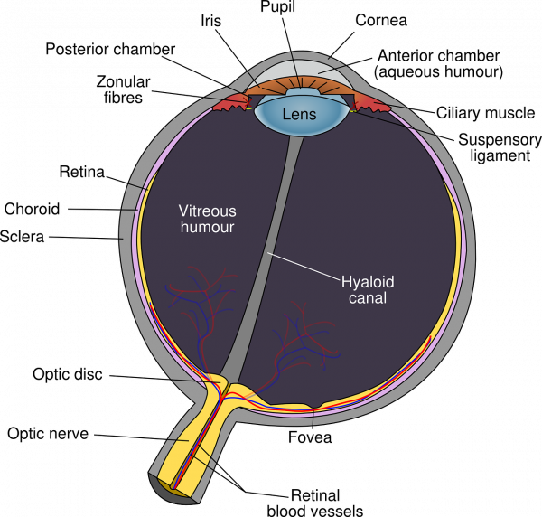
What we’re looking at during dilation
The lens
Cataracts are mostly a result of age or UV sun damage, however they can also be caused by trauma, medication, and certain health conditions. A dilated exam allows me to see your lenses more clearly, determining whether cataract is apparent, what type of cataract is present, and predicting the speed of progression so we can prepare you for surgery if necessary.
Retina and retinal blood vessels
Retinal blood vessels are assessed for the impact of high blood pressure, cholesterol, and diabetes. Bleeding and leakage of fats into the retina may be a sign of uncontrolled health conditions warranting a referral to your GP for further investigations.
Certain presentations of haemorrhaging can be a sign of leukaemia or cancer, as well as a result of vigorous activity or trauma.
Myopic (short-sighted) individuals are at higher risk of macula disease, and peripheral retina disease, showing as retinal thinning, increased adhesion, holes, or tears in the retina.
The macula is the central part of the eye that provides the highest resolution vision. This is the part we use for reading, or appreciating the finer details of faces and art.
Optic nerve
The optic nerve is the main structure we assess for potential brain tumours, which will cause the nerve to either swell or display paleness in colour. This is also where glaucoma takes place, elevated pressure within the eye causes thinning of the nerve fibre layer over time, leading to visual field loss.
If it’s been a while since your last comprehensive exam, or you’ve never had a dilated examination, book in and let’s take a proper look.

15 years of experience as an optometrist and owner of Carina Eye Care, experienced in prescribing the full range of myopia management options.
Written and medically reviewed by Dr Carina Trinh, BOptom · AHPRA Registered (OPT0001663121) · Optometry Australia Member · CCLSA Member · OSO Member · IOA Member.